DENTAL RADIOLOGY
X-RAY
Dental radiographs, commonly referred to as X-ray films, or informally X-rays, are formed by a controlled burst of X-ray radiation which penetrates oral structures at different levels, depending on varying anatomical densities, before striking the film or sensor. With the arrival of digital technology, which allows the reduction of doses and exposure time, the radiology has assumed an increasingly important role in the dental diagnostics.
The dental x-rays, which are most commonly performed in the dental practice, are:
• Periapical radiography - intraoral x-ray film of about 3 x 4 cm that allows seeing a complete tooth on the film, from the crown down to the tip of the root and surrounding tissues in their entirety. It allows a dentist to visualize abscess, cyst, tooth decay and is used as s "routine" x-ray during root canal treatment.
• Bitewing - intraoral x-ray film of about 3 x 4 cm that allows seeing some upper and lower teeth on the film. Routine bitewing radiographs are commonly used to examine for interdental caries and recurrent caries under existing restorations, as well as gum diseases.
• Panoramic X-ray - extraoral film that allows seeing all the upper and lower teeth along the jaw on a film size of 15 x 30 cm. It allows a quick way to get an overall view of an oral health and therefore is commonly used in the first visit.
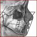
• Teleradiography - extraoral film of about 18 x 24 cm that allows seeing all the upper and lower teeth, the bones of the jaws and head and profile of the face in latero-lateral position. In dentistry it has been used particularly to plan orthodontic treatment.
• CT (computed tomography) - a medical imaging method that generates a three-dimensional (3D) image of an object. A large series of two-dimensional X-ray images are taken during rotation of a tube and a sensor around a patient. In dentistry it has been used particularly to plan dental implants, to detect the position of impacted teeth and nearby nerves. Specially designed CBCT (cone beam CT) scanners can be used instead, which produce adequate imaging with a tenfold reduction in radiation.
Dental X-rays are done in short time and saved on office computer. Patient receive duplicate in digital format.
MORE LINKS